Museum of the Human Inventions
Back to the collection

MHI 010 · Exhibition

The Microscope

c. 1590–1620

Disputed attribution

The Netherlands (Middelburg), with later advances in England and the Dutch Republic

Associated with

Zacharias Janssen and Hans Janssen
Spectacle-makers from Middelburg traditionally credited; the claim is uncertain
Hans Lippershey
Middelburg spectacle-maker; applied for a telescope patent in 1608
Robert Hooke
Published Micrographia in 1665; coined the term “cell”
Antonie van Leeuwenhoek
Made single-lens microscopes and observed bacteria and protozoa in the 1670s
Robert Koch
Used microscopy to identify the bacterium that causes tuberculosis in 1882
Plate for the exhibition on The Microscope
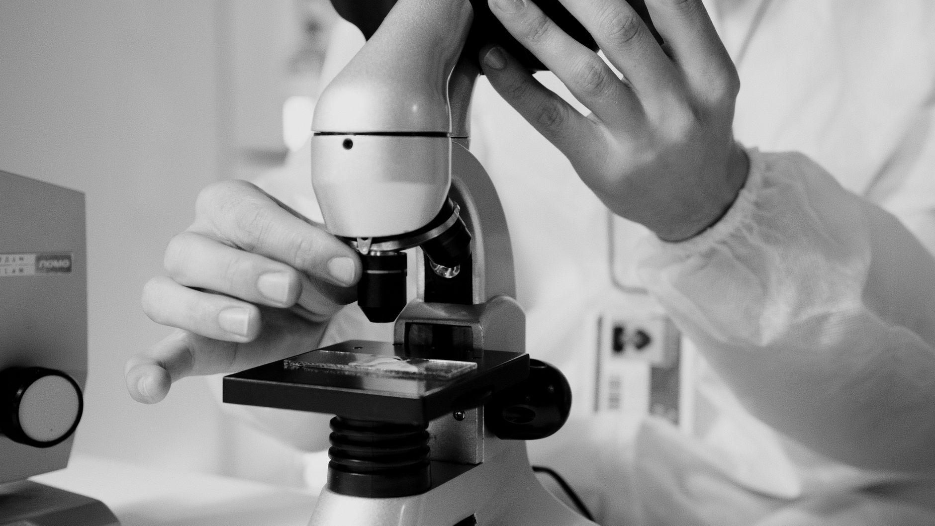
Illustrative photograph, not the original artifact. Photograph: Artem Podrez / Pexels

About this date

The compound microscope appeared in the Netherlands around the turn of the 17th century. Its exact date and inventor are uncertain.

About credit

Dutch spectacle-makers are credited with the first compound microscopes, but the Janssen claim rests on later testimony, and Lippershey and others have been named too. Historians treat the origin as uncertain.

Chapter I

The world before

The smallest things were unseen

People knew disease, decay and fermentation but could not see their causes. Explanations were drawn from tradition: imbalances of bodily humours, bad air, or divine judgement.

The smallest living things, and the structure of all living things, were beyond human sight.

The human achievement

Lenses, patience and curiosity

Spectacle-making had matured in Europe since around 1286, creating skilled lens grinders. In the Netherlands around 1600, combinations of lenses in a tube produced the first compound microscopes, though who made the first is uncertain.

In 1665, Robert Hooke published Micrographia, with engravings of fleas, fabric and a slice of cork, in which he described small chambers he called “cells”. In the 1670s, Antonie van Leeuwenhoek, a cloth merchant in Delft, ground tiny single lenses and reported seeing “animalcules” in rainwater, and in 1676, bacteria.

In the 19th century, Ernst Abbe and the Zeiss company improved optics through scientific design. Robert Koch used microscopes, stains and cultures to identify the bacteria behind tuberculosis (1882) and cholera (1883). The electron microscope, developed by Ernst Ruska and Max Knoll in 1931, reached much smaller scales.

Understanding the invention

How it works

A compound microscope magnifies in two stages.

Step 01 of 04

Light passes the specimen

A mirror or lamp lights a thin sample from below.

IV

The transformation

What changed for humanity

  1. Medicine

    Germ theory, developed through the work of Louis Pasteur, Robert Koch and others in the second half of the 19th century, changed hygiene, surgery and public health.

  2. Biology

    Cell theory in the 1830s–50s established the cell as the basic unit of life.

  3. Materials and industry

    Metallurgists, geologists and chip-makers inspect structures at tiny scales.

  4. Imagination

    Seeing a drop of water teeming with life changed how people understood their place in nature.

The microscope revealed cells, microbes and the structure of living things. It gave medicine and biology their foundations.

Before and after

Life before and after The Microscope

Before

Blamed on bad air or imbalances in the body.

After

Traced to specific microorganisms that can be seen, studied and targeted.

On larger screens, drag the divider (or use the arrow keys) to give more room to the world before or after.

VI

Its legacy

What came after

MHI 010

The Microscope

  • In this museum

    The Printing Press

    Printed, illustrated books such as Micrographia spread microscopic discoveries widely.

  • Beyond the collection

    Vaccines and antibiotics

    Both rely on understanding microbes first seen under a microscope.

  • Beyond the collection

    Genetics

    Chromosomes were first described through microscopy; DNA’s structure was found with related techniques.

  • Beyond the collection

    Semiconductor manufacturing

    Fabricating and inspecting chips depends on high-powered optics.

VII

Human stories

The people and the moments

A cloth merchant sees bacteria

Van Leeuwenhoek had no university training. He taught himself to grind lenses that magnified more than 200 times, and wrote to the Royal Society in London describing a world nobody had seen. At first some members doubted him; others later confirmed his observations.
Hooke’s cork

The “cells” Hooke saw were the walls of dead cork tissue, not living cells. The name stuck because they reminded him of monks’ rooms.

Where accounts differ, this museum follows the documented record and says so.

Built with